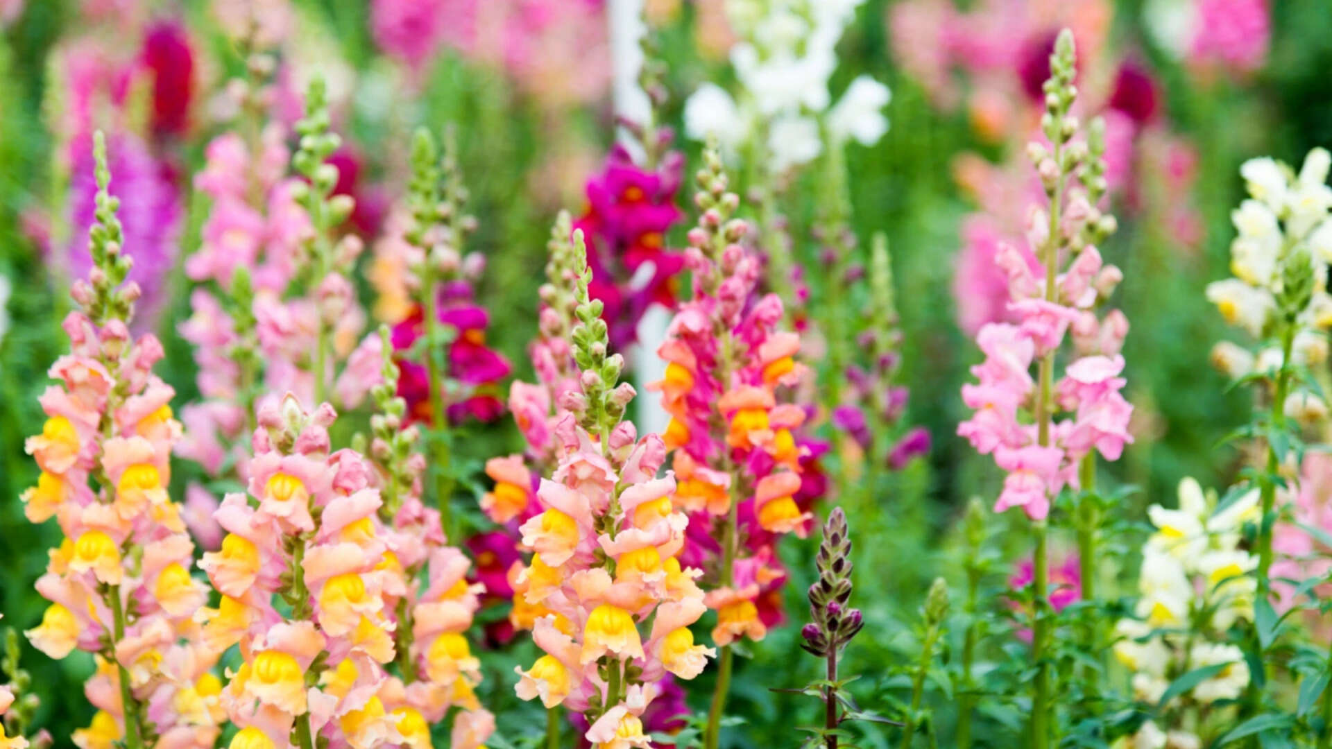
Floral phenotype divergence in wild snapdragons (Antirrhinum) operates as a high-precision signaling network optimized for hymenopteran visual systems. Rather than displaying random chromatic variation, Antirrhinum populations tune anthocyanin and carotenoid deposition along narrow geographic hybrid zones to balance opposing selective pressures: pollinator attraction efficiency versus reproductive isolation barriers. Understanding this signaling architecture requires mapping the biochemical synthesis pathways directly to the photoreceptor response curves of primary pollinators.
The Tri-Component Signaling Architecture
Visual communication in wild snapdragons relies on three distinct physiological inputs that map directly into the trichromatic visual space of Apis mellifera and Bombus species.
- Flavonoid Deposition Density: Anthocyanin accumulation—primarily cyanidin and delphinidin derivatives—determines the visual excitation of green (540 nm) and blue (440 nm) photoreceptors.
- Carotenoid Pigmentation Patterns: Aurone and lutein concentrations regulate ultraviolet absorbance and yellow reflectance, creating sharp visual contrast boundaries on the petal lobes.
- Epidermal Cell Geometry: Conical cell morphology on the adaxial petal surface acts as a physical light-focusing mechanism, increasing photon capture by internal pigment stores and reducing surface specular glare.
The primary objective of this architecture is maximizing signal contrast against a foliage background while minimizing energy expenditure in secondary metabolite synthesis.
Spectral Photoreceptor Mapping and Photonic Efficiency
Hymenopteran vision operates across three spectral peaks: ultraviolet (350 nm), blue (440 nm), and green (540 nm). Wild snapdragons engineer contrast through differential reflectance curves across these specific bandwidths.
Visual Space Delta = sqrt((Delta UV)^2 + (Delta Blue)^2 + (Delta Green)^2)
When Antirrhinum majus subspecies majus (magenta) and Antirrhinum majus subspecies striatum (yellow) interact along hybrid corridors, genetic loci control the precise spatial distribution of pigments.
Genetic Regulators of Pigment Synthesis
The transition between yellow and magenta phenotypes is governed by a small number of Mendelian loci with large phenotypic effects:
- ROSEA Locus: Encodes MYB-domain transcription factors (Rosea1 and Rosea2) that activate the anthocyanin biosynthetic pathway. Loss-of-function alleles suppress magenta pigmentation in the petal lobes.
- ELUTA Locus: Functions as a localized repressor of anthocyanin synthesis, restricting magenta coloration to specific regions such as the focal guides or lip.
- SULFUREA Locus: Regulates aurone (yellow pigment) accumulation. Recessive mutations abolish aurone synthesis, leaving the petals white in the absence of anthocyanins.
The epistatic interaction of ROSEA and SULFUREA dictates the precise coordinate within bee color space. A magenta flower produces high contrast in the blue-green photoreceptor channel against background foliage, while a yellow flower maximizes contrast in the UV-blue channel.
Hybrid Zone Dynamics and Evolutionary Cost Functions
In natural hybrid zones, such as those found across the Pyrenees, distinct color phenotypes exist in tight geographic clusters despite high rates of gene flow via wind and mobile pollinators. This persistence indicates strong frequency-dependent selection driven by pollinator behavior.
Bees exhibit strong flower constancy, systematically visiting the dominant floral phenotype within a foraging patch to minimize cognitive processing overhead. A rare variant within a dense patch suffers a fitness penalty due to reduced visitation rates and inefficient pollen transfer.
The Cost Function Matrix
- Low Frequency Phenotype Penalty: Decreased rate of cross-pollination due to foraging constancy of local pollinators.
- Metabolic Production Cost: High anthocyanin concentration requires significant resource allocation from the phenylpropanoid pathway, shifting carbon away from vegetative growth.
- Microclimate Vulnerability: Darker magenta petals absorb higher levels of solar radiation, raising internal flower temperature. While this can speed up pollen tube growth, it increases transpiration loss under drought stress.
This balance prevents any single phenotype from completely swamping adjacent populations, establishing stable hybrid boundaries that are maintained purely by visual signal processing dynamics.
Structural Petal Geometry as a Signal Amplifier
Pigmentation represents only half of the optical equation. The physical structure of the epidermal layer serves as a light-harvesting mechanism.
Conical epidermal cells act as micro-lenses. Light entering at an oblique angle is refracted downward into the interior mesophyll, increasing the probability of photon absorption by anthocyanin molecules stored in the cell vacuoles. This geometry reduces reflection off the outer cell wall, intensifying color saturation from various viewing angles.
When conical cell development is disrupted via mutations in the MIXTA gene, the petal surface flattens. Flat-celled petals exhibit high specular reflectance, appearing washed out under direct sunlight. Behavioral assays demonstrate that bees require significantly longer decision times to land on flat-celled mutants compared to wild-type conical-celled flowers, directly impacting foraging efficiency and floral reproductive throughput.
Strategic Deployment of Floral Guides
Spatial patterning on the floral lip serves as a physical landing target. Wild snapdragons concentrate aurones and anthocyanins around the entrance to the corolla tube, creating a steep visual gradient.
The lip pattern guides the bee directly to the mechanical opening mechanism. Antirrhinum flowers are closed structures requiring pollinators of a specific weight threshold—typically bumblebees—to force open the lower lip and access the nectar reward.
This mechanical filter ensures that visiting insects are effective vectors for pollen transfer rather than resource parasites. The visual guide reduces handling time per flower, increasing the net energy gain for the pollinator and maximizing the number of floral visits per foraging flight.
To optimize floral signaling models in experimental or agricultural environments, research strategies must prioritize calculating the exact perceptual distance within hymenopteran visual space rather than relying on human RGB measurements. Quantifying optical reflectance across 300-600 nm ranges combined with micro-CT scanning of epidermal cell structures provides the minimum viable dataset for predicting pollinator preference dynamics.